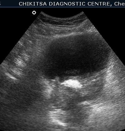
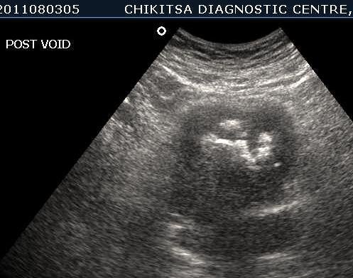
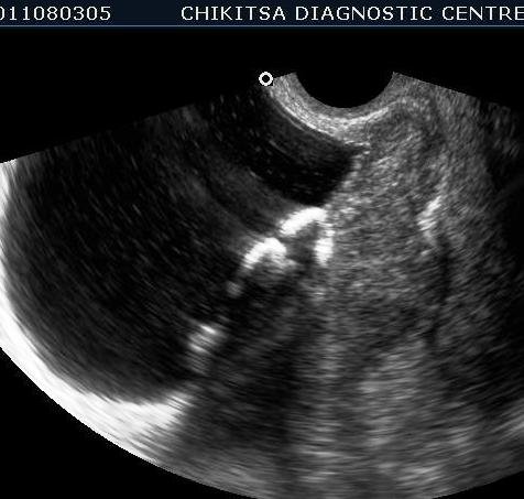
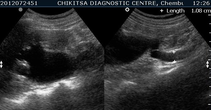
5 5 cm x 5 cm size ill-defined heterogeneous area in the posterior wall of the urinary bladder which is thickened and measures upto 0.6 cm in thickness. Large solid calcific areas are noted in the thicknened portion of the wall urinary bladder. Suggestive of schistosomiasis of the urinary bladder (possibly with malignant change)
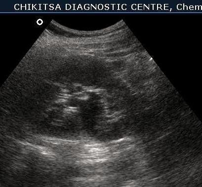
Left kidney shows Hydronephrosis
Left kidney shows Hydronephrosis
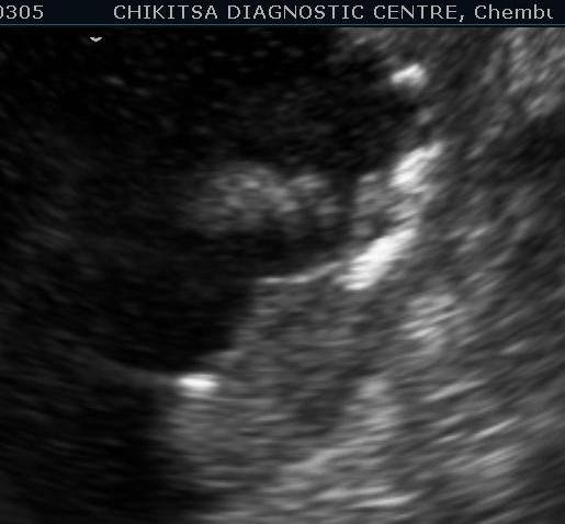
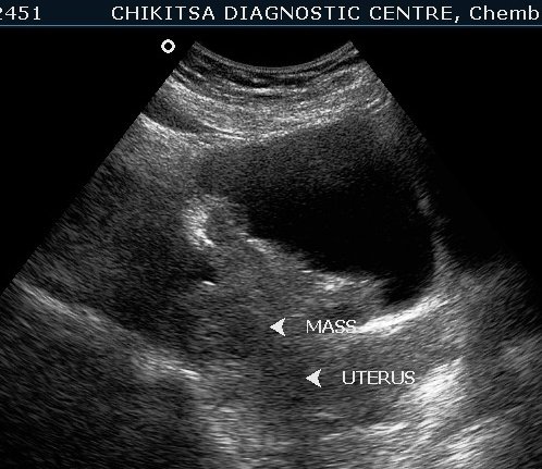
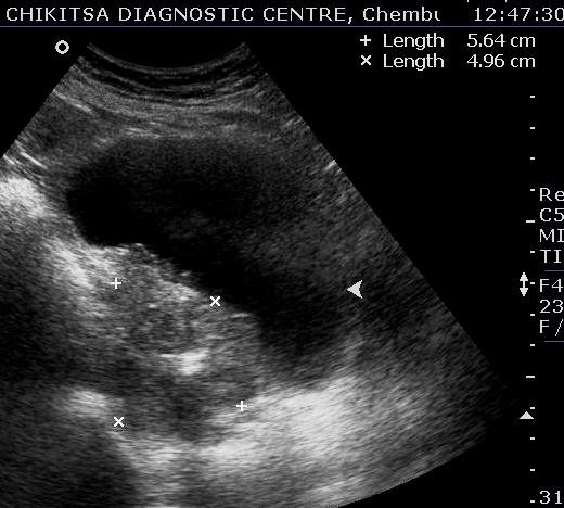
Lesion in the wall of the urinary bladder has increased in size. Calcific areas are noted. Urinary bladder malignancy ( confirmed by histopathology), possibly following schistosomiasis.
The lesion in the wall of the urinary bladder has increased in size. Calcific areas are noted. Urinary bladder malignancy ( confirmed by histopathology), possibly following schistosomiasis