What is Ovarian Reserve?
What is ovarian reserve? Learn about this key factor in female fertility, including static and dynamic tests like AMH, AFC, and FSH, and how it relates to age and reproductive potential.

★★★★★
4.6 – Ratings on Google
We Offer Comprehensive Ultrasound Training Programs covering Obstetrics, Gynecology, Fetal Echocardiography, Fetal Neurosonography, Doppler, Small Parts, Musculoskeletal Ultrasound, and More.
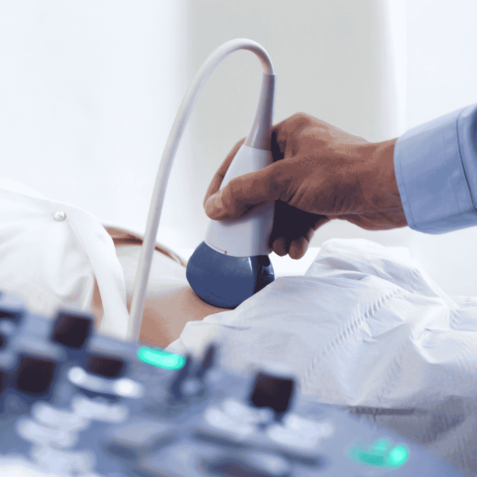
We offer specialized Sonography Orientations in Obstetrics ( including Doppler, 1st trimester screening, anomalies scan, fetal echocardiography, fetal neurosnography,3D/4D etc.), Gynecology (including infertility, fibroid mapping endometriosis mapping, urogynecology etc) Peripheral Doppler, Small parts, Musculoskeletal, NICU, IPCU, and Emergency Care Ultrasound, ensuring comprehensive training for diverse medical scenarios.
As a premier ultrasound training center in Mumbai and Maharashtra, we attract postgraduate doctors from across India, providing them with an unparalleled learning experience.
Our teaching methodology combines extensive hands-on ultrasound training with in-depth didactic lectures, ensuring a well-rounded understanding of the subject. We place a strong emphasis on academic excellence, encouraging our students to master both theoretical knowledge and practical application.
Beyond the classroom, we provide ongoing post-program support, guiding our students even after course completion.
Our faculty consists of highly experienced Radiologists with over 30 years of expertise in Sonography and Radiology, dedicated to delivering top-tier education and mentorship.
We offer comprehensive Training Programs that cover Basic and Advanced academic delivery in OBGY ultrasound and all other aspects of Ultrasound including Musculoskeletal, Peripheral Doppler, Small parts, NICU etc.
Ultrasound Fellowship Programs for Radiologists And OBGYs That Offer Specialized Training in all relevant Areas of Ultrasound With 6-month And 1-year options
Explore Diverse Ultrasound Applications Through Our Case Reports, Covering Pediatric GI, Thyroid Malignancy, Bronchial Atresia, Schistosomiasis, Syringomyelia, Mucinous Cystadenoma and More

What is ovarian reserve? Learn about this key factor in female fertility, including static and dynamic tests like AMH, AFC, and FSH, and how it relates to age and reproductive potential.

Understand ultrasound evaluation of LSCS (cesarean) scar sites. This module covers LUS thickness measurement techniques (transabdominal, transvaginal), timing, normal values, scar thinning, and the relationship between scar thickness and VBAC safety. Explore the challenges and current research in predicting uterine rupture risk.

Learn about the “Triangular Cord Sign,” a key ultrasound finding for diagnosing biliary atresia in infants. This module, contributed by Dr. Khatal and Dr. Badade, explains how this sign helps differentiate biliary atresia from neonatal hepatitis, emphasizing the importance of early diagnosis for successful Kasai procedures. Understand the sonographic appearance and anatomical basis of the triangular cord sign
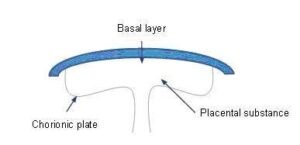
Learn placental grading (0-III) via ultrasound with Dr. Khatal. This module covers Grannum’s classification, correlating placental changes with gestational age, maternal health, and complications like IUGR. Explore placental calcification and its relationship with maternal age and parity

A 29-year-old female was presented with increased frequency of urination and tiny particles passing through urine.

A 28-year-old pregnant woman’s third-trimester ultrasound revealed findings suggestive of bronchial atresia. This case report details the ultrasound presentation, differential diagnosis, and postnatal characteristics of this rare congenital condition.