Master ultrasound with our training resources

comprehensive ultrasound training resources

We are passionate, patient, and effective communicators who break down complex problems into easy solutions, thus instilling enthusiasm and a better understanding of how to tackle various issues.

ultrasound training

What makes us special?

How do WE teach?

We specialize in adaptability and flexibility, ensuring every radiologist receives optimal support. Our training evolves with our students, continuously refining our methods to maximize their potential. Led by expert convenors, our clear, structured lessons simplify complex imaging concepts, reinforced with hands-on practical training. Our meticulously designed resources, developed in collaboration with leading radiologists, ensure relevance and real-world impact. With us, radiologists gain the skills and confidence to excel in their field.

Ultrasound Chikitsa Training Centre

Ultrasound Teaching Modules

Ultrasound Teaching Module

18 Feb 2025

What is Ovarian Reserve?

What is ovarian reserve? Learn about this key factor in female fertility, including static and dynamic tests like AMH, AFC, and FSH, and how it relates to age and reproductive potential.

Dr. Anirudh Badade

LSCS Scar 1

Ultrasound Teaching Module

18 Feb 2025

Ultrasound Evaluation Of The LSCS Scar Site

Understand ultrasound evaluation of LSCS (cesarean) scar sites. This module covers LUS thickness measurement techniques (transabdominal, transvaginal), timing, normal values, scar thinning, and the relationship between scar thickness and VBAC safety. Explore the challenges and current research in predicting uterine rupture risk.

Dr. Anirudh Badade

Triangular Cord Sign Of Biliary Atresia Ultrasound Module 1

Ultrasound Teaching Module

18 Feb 2025

Triangular Cord Sign Of Biliary Atresia

Learn about the "Triangular Cord Sign," a key ultrasound finding for diagnosing biliary atresia in infants. This module, contributed by Dr. Khatal and Dr. Badade, explains how this sign helps differentiate biliary atresia from neonatal hepatitis, emphasizing the importance of early diagnosis for successful Kasai procedures. Understand the sonographic appearance and anatomical basis of the triangular cord sign

Dr. Anirudh Badade

Placental Grading 1

Ultrasound Teaching Module

18 Feb 2025

Ultrasound Teaching Module on Placental Grading

Learn placental grading (0-III) via ultrasound with Dr. Khatal. This module covers Grannum's classification, correlating placental changes with gestational age, maternal health, and complications like IUGR. Explore placental calcification and its relationship with maternal age and parity

Dr. Anirudh Badade

Schistosomiasis of Urinary Bladder 1

Case Studies on Ultrasound & Sonography

17 Feb 2025

Schistosomiasis of Urinary Bladder : Ultrasound Case Study

A 29-year-old female was presented with increased frequency of urination and tiny particles passing through urine.

Dr. Anirudh Badade

Bronchial Atresia using Ultrasound 1

Case Studies on Ultrasound & Sonography

9 Feb 2025

Case Study on Bronchial Atresia using Ultrasound

A 28-year-old pregnant woman's third-trimester ultrasound revealed findings suggestive of bronchial atresia. This case report details the ultrasound presentation, differential diagnosis, and postnatal characteristics of this rare congenital condition.

Dr. Anirudh Badade

Case Studies on Ultrasound & Sonography

8 Feb 2025

Case Study on Thyroid Malignancy using Ultrasound

A 27-year-old female complained of neck swelling. Ultrasound and FNAC suggested follicular neoplasm. This case report discusses ultrasound features of thyroid malignancy and the importance of histopathology for diagnosis.

Dr. Anirudh Badade

Pediatric Gastrointestinal Ultrasound

Case Studies on Ultrasound & Sonography

7 Feb 2025

Case Study on Pediatric Gastrointestinal Ultrasound

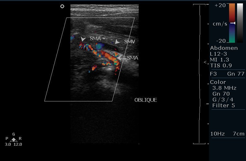
A 3-year-old boy with recurrent abdominal pain and vomiting was diagnosed with midgut volvulus, a complication of malrotation, using ultrasound. The "whirlpool sign," indicative of twisted intestines, was observed. This case highlights the importance of ultrasound in diagnosing this condition, especially in children with atypical presentations. Learn more about the clinical presentation, ultrasound findings, and surgical management of midgut volvulus.

Dr. Anirudh Badade

Learn how we've helped thousands of students achieve their academic dreams

Let's have a chat